brain stem
The brainstem interposes between the medulla and the diencephalon, lying ventrally to the cerebellum, that is, it connects the spinal cord with the brain structures located superiorly. The white matter of the brain stem includes tracts that receive and send motor and sensory information to the brain and also provide it.

his knowledge. Scattered in the white matter of the brainstem are masses of gray matter called nuclei, which exert intense effects on functions such as blood pressure and respiration. In its constitution enter bodies of neurons that are grouped in nuclei and nerve fibers, which in turn, are grouped in bundles called tracts, fascicles or lemnisci.
Many of the nuclei of the brainstem receive or imitate nerve fibers that enter into the constitution of the cranial nerves. Of the 12 pairs of cranial nerves, 10 connect to the brain stem.
 |  |  | ||
| Bulb | bridge | midbrain |
The brainstem is divided into: the medulla, situated caudally, the midbrain, and the pons located between them.
BULB (OBLONG MARROW) :
 - BRAIN STEM") |  - BRAIN STEM") |  - BRAIN STEM") |
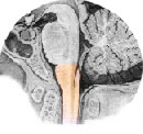
The medulla or medulla oblongata is shaped like a cone, the smaller end of which continues caudally with the spinal cord. As there is no line demarcating the separation between the medulla and the medulla, it is considered that the limit is in a horizontal plane that passes immediately above the most cranial root filament of the first cervical nerve, which corresponds to the level of the foramen magnum.
The upper limit of the bulb is made in a horizontal groove visible in the contour of this organ, the bulbopontine groove, which corresponds to the lower margin of the pons. The surface of the medulla is covered by two parallel grooves that continue into the medulla. These grooves delimit what is anterior and posterior in the medulla. Viewed from the surface, they appear as a continuation of spinal cord cords. The anterior median fissure ends cranially in a depression called the forme cecum.
On each side of the anterior median fissure is an eminence called the pyramid, formed by a compact bundle of descending nerve fibers that connect the motor areas of the brain to the motor neurons of the spinal cord. This tract is called the pyramidal tract or corticospinal tract.
In the caudal part of the medulla, the fibers of this tract obliquely cross the median plane and constitute the decussation of the pyramids. It is due to the decussation of the pyramids that the right cerebral hemisphere controls the left side of the body and the left cerebral hemisphere controls the right side. For example, in a right brain injury, the body will be affected in its entire left half.
| BULB (OBLONG MARROW) - BRAIN STEM - ANTERIOR VIEW |
 |
| Source: NETTER, Frank H.. Atlas of Human Anatomy. 2nd edition Porto Alegre: Artmed, 2000. |
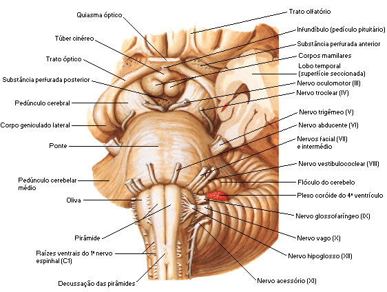
Between the anterior lateral and posterior lateral grooves we have the lateral area of the medulla, where an oval eminence, the olive, formed by a large amount of gray matter can be observed. Ventral to the olive, the reticular filaments of the hypoglossal nerve emerge from the anterior lateral sulcus. From the posterior lateral sulcus emerge the root filaments that unite to form the glossopharyngeal and vagus nerves, in addition to the filaments that constitute the cranial or bulbar root of the accessory nerve that unites with the spinal root.
| BULB (OBLONG MARROW) - BRAIN STEM - POSTERIOR VIEW |
 - BRAIN STEM - POSTERIOR VIEW") |
| Source: NETTER, Frank H.. Atlas of Human Anatomy. 2nd edition Porto Alegre: Artmed, 2000. |
The caudal half of the medulla or closed portion of the medulla is traversed by a narrow canal, a direct continuation of the central canal of the medulla, which opens to form the IV ventricle, whose floor is constituted by the rostral half or open portion of the medulla. The posterior median sulcus ends at mid-height of the medulla, due to the separation of its lips, which contribute to the formation of the lateral limits of the fourth ventricle.
Between the posterior median sulcus and the posterior lateral sulcus, there is the continuation of the posterior funiculus of the spinal cord, and in the medulla, this is divided into gracile fasciculus and cuneiform fasciculus by the posterior intermediate sulcus. These fascicles are made up of ascending nerve fibers from the spinal cord, which end in two masses of gray matter, the gracile and cuneiform nuclei, located in the most cranial part of the corresponding fascicles.
These nuclei determine the appearance of two eminences: the more medial gracile tubercle and the more lateral cuneiform tubercle. By virtue of the IV ventricle, the gracile and cuneiform tubercles depart laterally as two branches of a “V” and gradually continue upward with the inferior cerebellar peduncle (restiform body). This is formed by a thick bundle of fibers that form the lateral borders of the caudal half of the IV ventricle, flexing dorsally to penetrate the cerebellum.
| BULB (OBLONG MARROW) – BRAIN STEM – POSTERO-SIDE VIEW |
 - BRAIN STEM - POSTERO-SIDE VIEW") |
| Source: NETTER, Frank H.. Atlas of Human Anatomy. 2nd edition Porto Alegre: Artmed, 2000. |
The respiratory center is located in the medulla, which is very important for regulating the respiratory rhythm. The vasomotor center and the vomiting center are also located. The presence of the respiratory and vasomotor centers in the medulla makes lesions in this organ particularly dangerous.
Because of its importance in relation to vital functions, the medulla is often called the vital center. Because these structures are fundamental to the body, you can understand the seriousness of a fracture at the base of the skull. The bulb is also extremely sensitive to certain drugs, especially narcotics. An overdose of the narcotic causes bulb depression and death because the person stops breathing.
BRIDGE :
 |  |  |
Ponte is the part of the brainstem interposed between the medulla and the midbrain. It is situated ventrally to the cerebellum and rests on the basilar part of the occipital bone and the dorsum of the sella turcica of the sphenoid. Its ventrally located base presents a transverse striation due to the presence of numerous bundles of transverse fibers that run through it.
These fibers converge on each side to form a voluminous bundle, the middle cerebellar peduncle, which penetrates the corresponding cerebellar hemisphere. The point of emergence of the trigeminal nerve (cranial nerve V) is considered as the limit between the pons and the middle cerebellar peduncle (arm of the bridge). This emergence is made by two roots, a larger one, or sensory root of the trigeminal nerve, and a smaller one, or motor root of the trigeminal nerve.
| BRIDGE - BRAIN STEM - POSTERO-SIDE VIEW |
| |
| Source: NETTER, Frank H.. Atlas of Human Anatomy. 2nd edition Porto Alegre: Artmed, 2000. |
Running longitudinally along the ventral surface of the pons is a groove, the basilar groove, which usually houses the basilar artery.
The ventral part of the pons is separated from the medulla by the medullary groove.  pontine, from which the VI, VII and VIII cranial nerves emerge on each side from the midline.
The VI pair, the abducens nerve, emerges between the pons and the pyramid of the medulla. The eighth cranial nerve, the vestibulocochlear nerve, emerges laterally near a small lobe called a flocculus. The VII cranial pair, the facial nerve, emerges laterally with the VIII cranial pair, the vestibulocochlear nerve, with which it maintains intimate relations. Between the two, the intermediate nerve emerges, which is the sensory root of the VII cranial nerve.
| BRIDGE - BRAIN STEM - ANTERIOR VIEW |
| |
| Source: NETTER, Frank H.. Atlas of Human Anatomy. 2nd edition Porto Alegre: Artmed, 2000. |
The dorsal part of the pons does not present a line of demarcation with the dorsal part of the medulla, both constituting the floor of the fourth ventricle.
Bridge Cores :
![]() Trigeminal Nerve Motor Nucleus (V cranial nerve) – is situated on the lateral border of the fourth ventricle.
Trigeminal Nerve Motor Nucleus (V cranial nerve) – is situated on the lateral border of the fourth ventricle.
![]() Sensory Nuclei of the Trigeminal Nerve (V cranial nerve) – cephalic continuation of the sensory column of the spinal cord. Fibers entering the pons from the trigeminal ganglion divide into ascending and descending branches.
Sensory Nuclei of the Trigeminal Nerve (V cranial nerve) – cephalic continuation of the sensory column of the spinal cord. Fibers entering the pons from the trigeminal ganglion divide into ascending and descending branches.
![]() Abducens Nerve Nucleus (cranial nerve VI) – forms part of the dorsal gray matter of the medial eminence of the floor of the fourth ventricle, deep to the facial colliculus.
Abducens Nerve Nucleus (cranial nerve VI) – forms part of the dorsal gray matter of the medial eminence of the floor of the fourth ventricle, deep to the facial colliculus.
![]() Facial Nerve Nucleus (VII Cranial Pair) – is situated deep in the reticular formation, lateral to the abducens nerve nucleus. They emerge at the edge of the caudal between the olive and the inferior cerebellar peduncle.
Facial Nerve Nucleus (VII Cranial Pair) – is situated deep in the reticular formation, lateral to the abducens nerve nucleus. They emerge at the edge of the caudal between the olive and the inferior cerebellar peduncle.
![]() Vestibulocochlear Nerve Nucleus (cranial nerve VIII) – the nucleus of the vestibular division occupies a large area in the lateral portion of the fourth ventricle. The nucleus of the cochlear division is located in the caudal portion of the pons.
Vestibulocochlear Nerve Nucleus (cranial nerve VIII) – the nucleus of the vestibular division occupies a large area in the lateral portion of the fourth ventricle. The nucleus of the cochlear division is located in the caudal portion of the pons.
| BRIDGE - SCHEME OF BRIDGE NUCLEUS - BRAIN STEM - SIDE VIEW |
 |
Source: NETTER, Frank H.. Atlas of Human Anatomy. 2nd edition Porto Alegre: Artmed, 2000. |
Fourth Ventricle : It is located between the medulla and the pons on its posterior surface and ventrally to the cerebellum. It continues caudally with the central canal of the medulla and cranially with the cerebral aqueduct, the midbrain cavity that communicates the III and IV ventricles. The cavity of the fourth ventricle extends on each side to form the lateral recesses situated on the dorsal surface of the inferior cerebellar peduncle.  This recess communicates on each side with the subarachnoid space through the two lateral openings of the fourth ventricle. There is also a median opening of the fourth ventricle called the form of Magendie, or median foramen, situated in the middle of the caudal half of the roof of the fourth ventricle. Through this cavity, the cerebrospinal fluid, which fills the ventricular cavity, passes into the subarachnoid space.
| BRIDGE - FOURTH VENTRICLE - BRAIN STEM - SIDE VIEW |
 |
| Source: NETTER, Frank H.. Atlas of Human Anatomy. 2nd edition Porto Alegre: Artmed, 2000. |
The floor of the IV ventricle, or rhomboid fossa, is formed by the dorsal part of the pons and the open portion of the medulla.
IV Ventricle Roof : The cranial half of the IV ventricle roof is made up of a thin sheet of white matter, the superior medullary velum, which extends between the two superior cerebellar peduncles. In the constitution of the caudal half we have the following formations:
![]() A small part of the white matter of the cerebellar nodule.
A small part of the white matter of the cerebellar nodule.
![]() The inferior medullary velum, bilateral formation consisting of a thin white sheet attached medially to the lateral edges of the cerebellar nodule.
The inferior medullary velum, bilateral formation consisting of a thin white sheet attached medially to the lateral edges of the cerebellar nodule.
![]() Choroid screen of the fourth ventricle, which joins the two anterior formations to the edges of the caudal half of the floor of the fourth ventricle.
Choroid screen of the fourth ventricle, which joins the two anterior formations to the edges of the caudal half of the floor of the fourth ventricle.
| BRIDGE - FOURTH VENTRICLE FLOOR - BRAIN STEM - POSTERIOR VIEW |
| |
| Source: NETTER, Frank H.. Atlas of Human Anatomy. 2nd edition Porto Alegre: Artmed, 2000. |
The choroidal screen is formed by the union of the ependymal epithelium, which internally lines the ventricle with the pia mater and externally reinforces this epithelium. This screen emits irregular and highly vascularized projections for the formation of the choroid plexus of the fourth ventricle. This choroid plexus is T-shaped and produces cerebrospinal fluid, which accumulates in the ventricular cavity passing into the subarachnoid space through the lateral and median openings of the fourth ventricle.
The pons plays a key role in regulating the breathing pattern and rhythm. Injuries to this structure can cause serious disturbances in respiratory rhythm.
midbrain :
 |  |  |
It interposes between the pons and the brain, of which it is represented by a plane that connects the two mammillary bodies, belonging to the diencephalon, to the posterior commissure. It is crossed by a narrow channel, the cerebral aqueduct. The part of the midbrain situated dorsal to the aqueduct is the roof of the midbrain. Ventrally, we have the two cerebral peduncles, which in turn are divided into a dorsal part, the tegmentum, and a ventral part, the base of the peduncle.
 In a cross-section of the midbrain, the tegmentum is seen to be separated from the base by a dark area, the substantia nigra (nigra). Next to the substantia nigra there are two longitudinal grooves: one lateral, the lateral sulcus of the midbrain, and the other medial, the medial sulcus of the cerebral peduncle. These grooves mark the boundary between the base and the tegmentum of the cerebral peduncle. From the medial sulcus emerges the oculomotor nerve, 3rd cranial nerve.
Midbrain roof: in dorsal view, the midbrain roof has four rounded eminences called superior and inferior colliculi, separated by two perpendicular cross-shaped grooves. In the anterior part of the longitudinal branch of the cross, the pineal body, which belongs to the diencephalon, is housed. Caudally to each inferior colliculus emerges the fourth cranial nerve, the trochlear nerve.
| MISENCEPHALO (DIDACTIC SCHEME) – CROSS-SECTION OF THE MISENCEPHALO |
 - CROSS-SECTION OF THE MISENCEPHALO") |
| Source: MACHADO, Ângelo. Functional Neuroanatomy. Rio de Janeiro/São Paulo: Atheneu, 1991. |
Each colliculus attaches to a small oval eminence of the diencephalon, the geniculate body, through a superficial bundle of nerve fibers that constitutes its arm. Thus, the inferior colliculus attaches to the medial geniculate body by the inferior colliculus arm, and the superior colliculus attaches to the lateral geniculate body by the superior colliculus arm, which has its hidden path between the pulvinar of the thalamus and the medial geniculate body. The lateral geniculate body is found at the end of the optic tract.
| MISENCEPHALO – COLLICULUS AND GENICULATE BODIES – POSTERO-LATERAL VIEW |
| |
| Source: NETTER, Frank H.. Atlas of Human Anatomy. 2nd edition Porto Alegre: Artmed, 2000. |
Cerebral Peduncles: Viewed ventrally, the cerebral peduncles appear with two large fiber bundles that arise at the upper edge of the pons and diverge cranially to penetrate deep into the brain. Thus, they delimit a deep triangular depression, the interpeduncular fossa, limited anteriorly by two eminences belonging to the diencephalon, the mammillary bodies. The bottom of the interpeduncular fossa has small holes for the passage of vessels. It is called posterior perforated substance.
Red nucleus – occupies a large part of the tegmentum. It is an oval-shaped mass that extends from the caudal limit of the superior colliculus to the subthalamic region. It is circular in cross section.
| LOWER VIEW OF THE MEDBRANE |
 |
| Source: NETTER, Frank H.. Atlas of Human Anatomy. 2nd edition Porto Alegre: Artmed, 2000. |
Midbrain Nuclei :
![]() Nucleus of the Mesencephalic Root of the Trigeminal Nerve (cranial nerve V) – forms a dispersed region in the lateral portion of the central gray matter that surrounds the aqueduct.
Nucleus of the Mesencephalic Root of the Trigeminal Nerve (cranial nerve V) – forms a dispersed region in the lateral portion of the central gray matter that surrounds the aqueduct.
![]() Trochlear Nerve Nucleus (cranial nerve IV) – is at the level of the inferior colliculus.
Trochlear Nerve Nucleus (cranial nerve IV) – is at the level of the inferior colliculus.
![]() Oculomotor Nerve Nucleus (3rd cranial nerve) – appears in cross section. It extends to the superior colliculus.
Oculomotor Nerve Nucleus (3rd cranial nerve) – appears in cross section. It extends to the superior colliculus.
| MIDBRAIN - NUCLEUS SCHEME - BRAIN STEM - POSTERIOR VIEW |
 |
| Source: NETTER, Frank H.. Atlas of Human Anatomy. 2nd edition Porto Alegre: Artmed, 2000. |
Cerebellar Peduncles Review:
![]() Inferior cerebellar peduncle: originates from the medulla.
Inferior cerebellar peduncle: originates from the medulla.
![]() Middle Cerebellar Peduncle: Originates from the pons.
Middle Cerebellar Peduncle: Originates from the pons.
![]() Superior Cerebellar Peduncle: Originates from the midbrain.
Superior Cerebellar Peduncle: Originates from the midbrain.
| Inferior cerebellar peduncle Origin: bulb | Middle Cerebellar Peduncle Origin: Bridge | Superior Cerebellar Peduncle Origin: Midbrain | ||
 |  |  |