Nervous system
During the evolution of the living being, we saw that the first neurons appeared on the external surface of the organism, considering that the primary function of the nervous system is to relate the animal to the environment. Of the three embryonic layers, the ectoderm is the one that is in contact with the external environment of the organism and it is from this layer that the nervous system originates.
The first index  The process of formation of the nervous system consists of a thickening of the ectoderm, situated above the notochord, forming the so-called neural plate. It is known that the formation of this plate and the subsequent formation of the neural tube plays an important role in the inducing action of the notochord and mesoderm. Notochords implanted in the abdominal wall of amphibian embryos induce neural tube formation there. Notochord or mesoderm extirpations in young embryos resulted in large marrow anomalies.
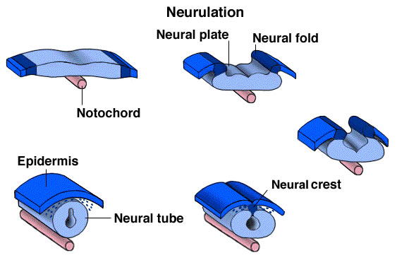
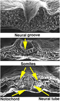
The neural plate grows progressively, becomes thicker, and acquires a longitudinal groove called the neural groove that deepens to form the neural groove. The lips of the neural gutter fuse to form the neural tube.
The undifferentiated ectoderm then closes over the neural tube, thus isolating it from the external environment. At the point where this ectoderm meets the lips of the neural groove, cells develop that form on each side a longitudinal lamina called the neural crest. The neural tube gives rise to elements of the central nervous system, while the crest gives rise to elements of the peripheral nervous system, in addition to elements not belonging to the nervous system.
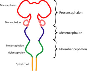
From the beginning of its formation, the diameter of the neural tube is not uniform. The cranial part, which gives rise to the adult brain, becomes dilated and constitutes the primitive brain, or archencephalon; the caudal part, which gives rise to the adult medulla, remains with a uniform caliber and constitutes the primitive medulla of the embryo. In the archencephalon, three dilatations are initially distinguished, which are the primordial encephalic vesicles called: forebrain, midbrain and rhombencephalon. With the subsequent development of the embryo, the forebrain gives rise to two vesicles, telencephalon and diencephalon. The midbrain does not change, and the rhomboencephalon gives rise to the metencephalon and myelocephalon.
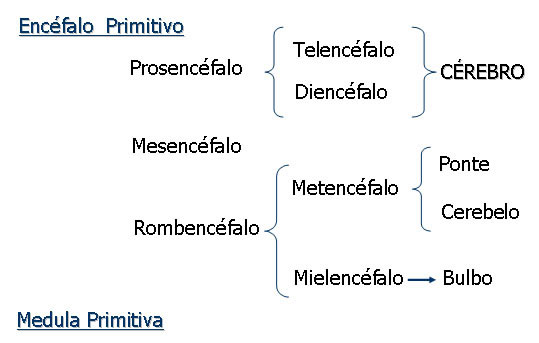

The telencephalon comprises a medial part, from which two lateral portions, the lateral telencephalic vesicles, envaginate. The middle part is closed anteriorly by a lamina that constitutes the most cranial portion of the nervous system and is called lamina terminalis. The lateral telencephalic vesicles grow large to form the cerebral hemispheres and almost completely hide the midbrain and diencephalon.
The diencephalon has four small diverticula: two lateral, the optic vesicles, which form the retina; a dorsal, which forms the pineal gland; and a ventral one, the infundibulum, which forms the neurohypophysis.
Neural Tube Cavity : The neural tube lumen remains in the adult nervous system, undergoing, in some parts, several modifications. The lumen of the primitive medulla forms, in the adult, the central canal of the medulla. The dilated cavity of the hindbrain forms the fourth ventricle. The cavity of the diencephalon and the middle part of the telencephalon form the third ventricle.
The midbrain lumen remains narrow and forms the cerebral aqueduct that joins the III to the IV ventricle. The lumen of the lateral telencephalic vesicles form, on each side, the lateral ventricles, joined to the third ventricle by the two interventricular foramina. All cavities are lined by a cuboidal epithelium called the ependyma and, with the exception of the central canal of the spinal cord, contain a cerebrospinal fluid, or CSF.
Flexures : during the development of the different parts of the archencephalon, flexures or curvatures appear in its ceiling or floor, mainly due to different growth rhythms. The first flexure to appear is the cephalic flexure, which appears in the region between the midbrain and forebrain. Soon, between the primitive medulla and the archencephalon, a second flexure appears, called the cervical flexure. It is determined by ventral flexion of the entire head of the embryo in the region of the future neck. Finally, a third flexure appears, opposite the first two, at the point of union between the meta and the myelencephalon: the pontine flexure. With development, the two caudal flexures dissolve and practically disappear. However, the cephalic flexure remains determined, in the adult male brain, an angle between the brain, deriving from the forebrain, and the rest of the neuraxis.
Division of the nervous system based on anatomical and functional criteria

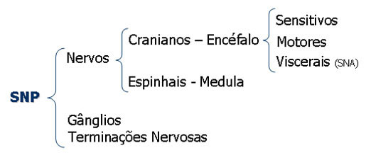
The central nervous system is the one located within the axial skeleton (cranial cavity and vertebral canal); the peripheral nervous system is the one that is located outside this skeleton. The brain is the part of the central nervous system situated within the neural skull; and the medulla is located within the vertebral canal. The brain and spinal cord constitute the neuro-axis. In the brain we have the cerebrum, cerebellum and brain stem.

The nervous system can be divided into the nervous system of the relational life, or somatic, and the nervous system of the vegetative, or visceral life. The nervous system of relationship life is the one that relates the organism with the environment. It has an afferent and an efferent component.
The afferent component conducts impulses originating in peripheral receptors to the nerve centers, informing them about what is happening in the environment. The efferent component leads to the skeletal striated muscles the command of the nerve centers resulting in voluntary movements.
The visceral nervous system is the one that is related to the innervation and control of the viscera. The afferent component carries nerve impulses originating from receptors in the viscera to specific areas of the nervous system. The efferent component carries impulses originating in nerve centers to the viscera. This efferent component is also called the autonomic nervous system and can be divided into sympathetic and parasympathetic nervous systems.
