Uterine tubes
Fallopian tube is a paired tube that is implanted on each side at the respective latero-superior angle of the uterus, and projects laterally, representing the horizontal branches of the tube.
This tube is irregular in terms of caliber, presenting approximately 10 cm in length.
It dilates as it moves away from the uterus, opening distally through a true funnel with a fringed edge.
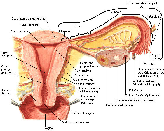
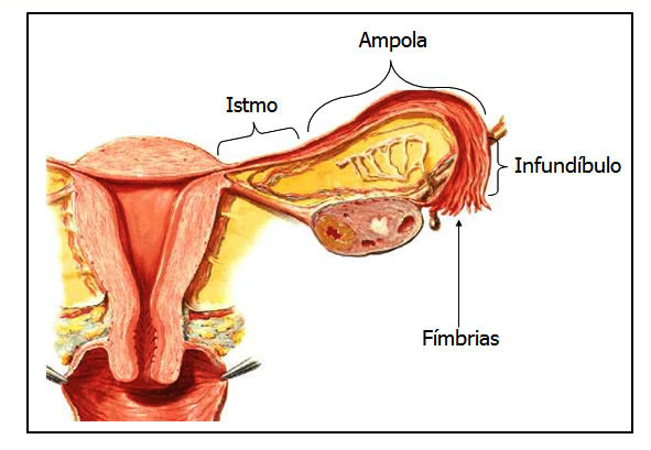
The fallopian tube is divided into 4 regions, which in the mediolateral direction are: Uterine Part, Isthmus, Ampule and Infundibulum .
The Uterine Part is the intramural portion, that is, it constitutes the segment of the tube that is located in the wall of the uterus.
At the beginning of this portion of the tube, we find an orifice called the uterine ostium of the tube, which establishes its communication with the uterine cavity.
The isthmus is the less caliber portion, located next to the uterus, while the ampulla is the dilatation that follows the isthmus.
The ampoule is considered the place where the fertilization of the egg by the sperm is normally processed.
The most distal portion of the tube is the Infundibulum , which can be compared to a funnel whose mouth has a very irregular edge, taking on the appearance of fringes.
These fringes are called tuba fimbriae, one of which stands out for being longer, called the ovarian fimbria.
The infundibulum opens freely into the peritoneal cavity through a foramen known as the uterine tube abdominal ostium.
The horizontal part would be represented by the isthmus and the vertical by the ampulla and infundibulum.
Commonly, the infundibulum fits over the ovary, and the fimbriae could be roughly compared to the fingers of a hand holding an orange above.
Structurally, the fallopian tube is constituted by four concentric layers of tissues that are, from the periphery to the depth, the serous tunic, subserous mesh, muscular tunic and mucous tunic.
The muscular coat, represented by smooth muscle fibers, allows peristaltic movements to the tube, helping the migration of the ovum towards the uterus.
The mucous membrane is formed by hair cells and has numerous longitudinal parallel folds, called tubal folds.
The fallopian tube has two functions:
- Transport the egg from the ovary to the uterus;
- Place where fertilization of the egg by the sperm takes place.
| DIVISIONS OF THE UTERINE TUBE |
 |
| Source: NETTER, Frank H.. Atlas of Human Anatomy. 2nd edition Porto Alegre: Artmed, 2000. |

