Abdominal Muscles
The abdominal muscles can be divided into 4 regions:
- Anterolateral Region
- posterior region
- Upper Region
- Lower Region
| Anterolateral Region | posterior region | |
| Anterior Rectus Abdomen abdomen pyramid External Abdominal Oblique Internal oblique of the abdomen transversus abdominis | Lumbar Square iliopsoas Psoas Minor | |
| Upper Region | Lower Region | |
| Diaphragm | levator ani ischiococcygeus |
Anterolateral Region
1. ANTERIOR ABDOMINAL RECTUM
Superior Insertion : External and inferior surface of 5th to 7th costal cartilages and xiphoid process
Inferior Insertion : Body of pubis and pubic symphysis
Innervation : Last 5 intercostal nerves
Action : Increased intra-abdominal pressure (Expiration, Vomiting, Defecation, Urination and Childbirth)
* Fixed in the Chest : Retroversion of the pelvis
* Fixed in the Pelvis : Flexion of the trunk (+ or – 30°)
2. ABDOMINAL PYRAMID
Upper Insert : Alba Line
Inferior Insertion : Body of pubis and anterior pubic ligament
Innervation : 12th intercostal nerve
Action : Tense the linea alba
3. EXTERNAL ABDOMINAL OBLIQUE
Superior Insertion : External surface of last 7 ribs
Inferior Insertion : Anterior ½ of the iliac crest, ASIS, pubic tubercle and linea alba
Innervation : last 4 intercostal nerves, iliohypogastric and ilioinguinal nerves
Action :
* Unilateral Contraction : Rotation with chest rotating to the opposite side
* Bilateral contraction : Flexion of the trunk and increase in intra-abdominal pressure
4. INTERNAL ABDOMEN OBLIQUE
Superior Insertion : Last 3 costal cartilages, pubic crest and linea alba
Inferior Insertion : Iliac crest, ASIS, and inguinal ligament
Innervation : last 4 intercostal nerves, iliohypogastric and ilioinguinal nerves
Action : Same as the External Oblique, but rotates the thorax to the same side.
5. TRANSVERSE ABDOMEN
Posterior Insertion : Inner surface of last 6 costal cartilages, thoracolumbar fascia, iliac crest, and inguinal ligament
Anterior Insertion : Linea alba and pubic crest
Innervation : 5 last intercostals, iliohypogastric and ilioinguinal nerves
Action : Increase intra-abdominal pressure and stabilization of the lumbar spine
posterior region
1. LUMBAR SQUARE
Superior Insertion : 12th rib and transverse process of 1st to 4th lumbar vertebrae
Inferior Insertion : Iliac crest and ileolumbar ligament
Innervation : 12th intercostal nerve and L1
Action : Homolateral trunk tilt and 12th rib depression
2. ILIOPSOAS
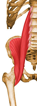 Iliac
Iliac
Superior Insertion : Superior 2/3 of the iliac fossa, iliac crest and sacral wing
Lower Insertion : Lesser Trochanter
Innervation : Femoral Nerve (L2 – L3)
Action : Hip flexion, pelvis anteversion and lumbar spine flexion (30° – 90°)
psoas major
Superior Insertion : Transverse process of lumbar vertebrae, intervertebral bodies and discs of the last thoracic and all lumbar
Lower Insertion : Lesser Trochanter
Innervation : Superior and inferior nerve of the psoas major muscle (L1 – L3)
Action : Thigh flexion, lumbar spine flexion (30° – 90°) and homolateral bending
3. PSOAS MINOR (It is usually absent)
Superior Insertion : Vertebral body of T12 and L1
Lower Insertion : Iliopectineal Eminence
Innervation : L1
Action : Flexion of the pelvis and lumbar spine
Upper Region
1. DIAPHRAGM
Origin : Inner surface of the last 6 ribs, inner surface of the xiphoid process and vertebral bodies of the upper lumbar vertebrae
Insertion : In the central tendon (aponeurosis)
Innervation : Phrenic nerve (C3 – C5) and last 6 intercostal nerves (proprioception)
Action : Inspiratory, as it reduces the internal pressure of the rib cage allowing air to enter the lungs, stabilization of the spine and expulsions (defecation, vomiting, urination and delivery)
Lower Region
1. ANAL LIFTER
The levator ani usually shows a two-part separation:
– Pubococcygeus
– Iliococcygeus
Origin : Between the superior ramus of the pubis and the ischial spine
Insertion : Coccyx, sphincter of anus and at the central tendon point of the perineum
Innervation : Pudendal Plexus (S3 - S5)
Action : Supports and slightly elevates the pelvic floor, resisting increased intra-abdominal pressure, such as during forced expiration
2. ISCHIOCOCCYGEOUS
Origin : Apex of ischial spine and sacrospinous ligament
Insertion : Margin of the coccyx and on the lateral surface of the sacrum
Innervation : Pudendal Plexus (S4 - S5)
Action : Pulls the coccyx ventrally, supporting the pelvic floor against intra-abdominal pressure
| ABDOMINAL MUSCLES Previous View - Surface Layer |
 |
| Source: NETTER, Frank H.. Atlas of Human Anatomy. 2nd edition Porto Alegre: Artmed, 2000 . |
| ABDOMINAL MUSCLES Previous View - Middle Layer |
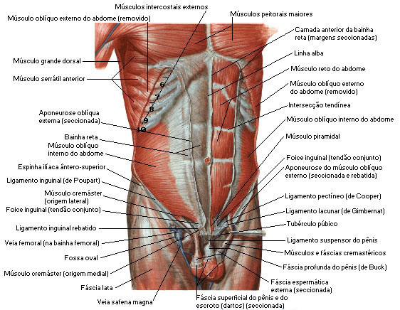 |
| Source: NETTER, Frank H.. Atlas of Human Anatomy. 2nd edition Porto Alegre: Artmed, 2000. |
| ABDOMINAL MUSCLES Previous View - Deep Layer |
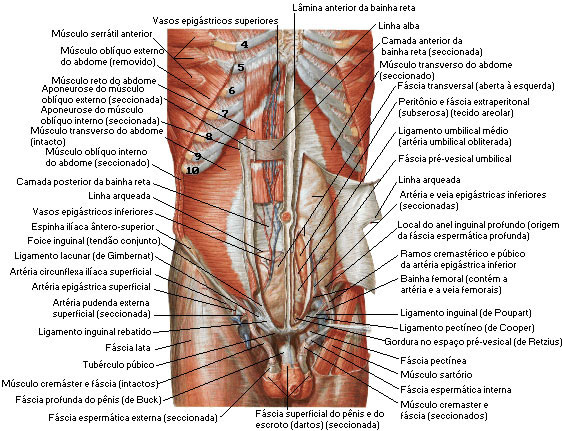 |
| Source: NETTER, Frank H.. Atlas of Human Anatomy. 2nd edition Porto Alegre: Artmed, 2000. |
| ABDOMINAL MUSCLES iliopsoas |
 |
| Source: NETTER, Frank H.. Atlas of Human Anatomy. 2nd edition Porto Alegre: Artmed, 2000. |
| ABDOMINAL MUSCLES Iliopsoas action |
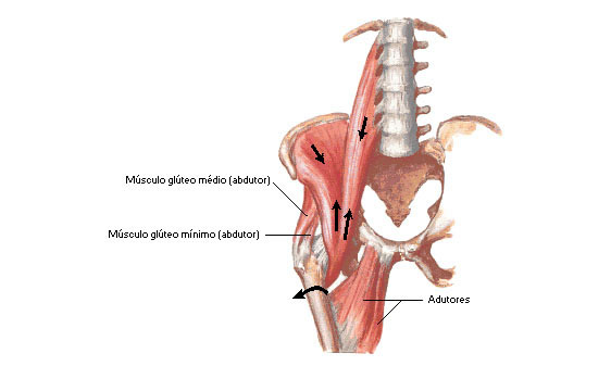 |
| Source: NETTER, Frank H.. Atlas of Human Anatomy. 2nd edition Porto Alegre: Artmed, 2000. |
| ABDOMINAL MUSCLES Internal View of the Posterior Abdominal Wall |
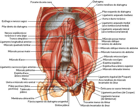 |
| Source: NETTER, Frank H.. Atlas of Human Anatomy . 2nd edition Porto Alegre: Artmed, 2000. |
