Cervical Plexus
Formed by the ventral branches of the four upper cervical nerves, it innervates some muscles in the neck, the diaphragm, and areas of skin in the head, neck, and chest.
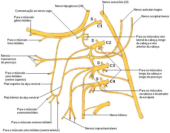
Each ventral branch anastomoses with the subsequent one, forming three loops of lateral convexity (C1 with C2, C2 with C3 and C3 with C4). From these three loops derive branches that constitute the two parts of the cervical plexus (superficial and deep).
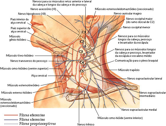
The superficial part consists of essentially sensory fibers, which form a bundle that appears at the level of the middle of the posterior border of the sternocleidomastoid muscle, at which point the fillets fan out to the skin in the surrounding region, to the pinna, to the skin. neck and the region close to the collarbone.
The deep part of the plexus is constituted by motor fibers, destined to the anterolateral musculature of the neck and the diaphragm. For this, in addition to branches that leave the three loops separately, we find two important formations, which are the cervical loop and the phrenic nerve.
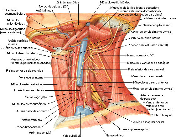
The cervical loop is formed by two roots, one superior and one inferior. The superior root of the ansa cervicalis reaches the hypoglossal nerve as it descends in the neck. The lower root descends a few centimeters laterally to the internal jugular vein, then curves forward, anastomosing with the upper root.
The ansa cervicalis gives off branches that innervate all the infrahyoid muscles.
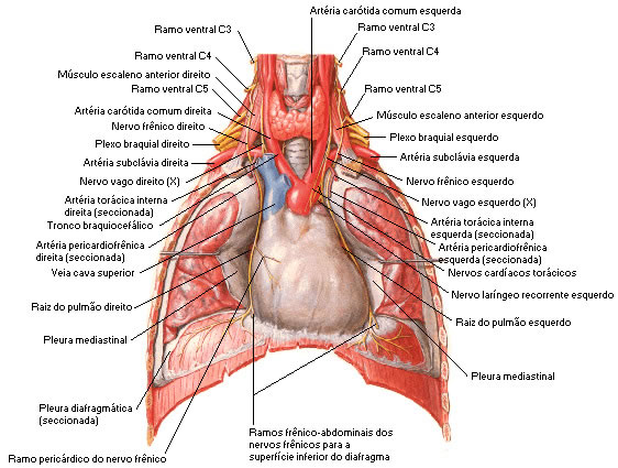
The phrenic nerve, formed by motor fibers that derive from C3, C4 and C5, descends in front of the anterior scalene muscle, passes along the pericardium, to distribute itself in the diaphragm.
Each branch, except the first, divides into ascending and descending parts that unite in communicating loops. From the first loop (C2 and C3) arise superficial branches that innervate the head and neck; from the second loop (C3 and C4) the cutaneous nerves of the shoulder and thorax originate. Branches are superficial or deep; the superficial branches pierce the cervical fascia to innervate the skin, while the deep branches innervate the muscles.
The superficial branches form ascending and descending groups and the deep series medial and lateral.
Ascending Surfaces :
![]() Lesser Occipital Nerve (C2) – innervates the skin of the region posterior to the pinna;
Lesser Occipital Nerve (C2) – innervates the skin of the region posterior to the pinna;
| MINOR OCCIPITAL NERVE |
 |
| Source: NETTER, Frank H.. Atlas of Human Anatomy. 2nd edition Porto Alegre: Artmed, 2000. |
![]() Great Auricular Nerve (C2 and C3) – its anterior branch innervates the skin of the face over the parotid gland, communicating with the facial nerve; the posterior branch supplies the skin over the mastoid process and over the back of the pinna;
Great Auricular Nerve (C2 and C3) – its anterior branch innervates the skin of the face over the parotid gland, communicating with the facial nerve; the posterior branch supplies the skin over the mastoid process and over the back of the pinna;
![]() Transverse Neck Nerve (C2 and C3) – its ascending branches ascend to the submandibular region, forming a plexus with the cervical branch of the facial nerve below the platysma; the descending branches pierce the platysma and are distributed anterolaterally to the skin of the neck, down to the lower part of the sternum.
Transverse Neck Nerve (C2 and C3) – its ascending branches ascend to the submandibular region, forming a plexus with the cervical branch of the facial nerve below the platysma; the descending branches pierce the platysma and are distributed anterolaterally to the skin of the neck, down to the lower part of the sternum.
Descending Surfaces :
![]() Medial Supraclavicular Nerves (C3 and C4) – innervate the skin up to the midline, lower part of the second rib and the sternoclavicular joint;
Medial Supraclavicular Nerves (C3 and C4) – innervate the skin up to the midline, lower part of the second rib and the sternoclavicular joint;
![]() Intermediate Supraclavicular Nerves – innervate the skin over the pectoralis major and deltoid muscles along the level of the second rib;
Intermediate Supraclavicular Nerves – innervate the skin over the pectoralis major and deltoid muscles along the level of the second rib;
![]() Lateral Supraclavicular Nerves – innervate the skin of the upper and posterior parts of the shoulder.
Lateral Supraclavicular Nerves – innervate the skin of the upper and posterior parts of the shoulder.
Deep Branches – Medial Series :
Branches communicating with the hypoglossal, vague and sympathetic; the muscular branches innervate the rectus capitis lateralis (C1), anterior rectus capitis (C1 and C2), longus capitis (C1, C2 and C3), longus neck (C2-C4), inferior root of the cervical loop (C1 and C2). C2-C3), infrahyoid muscles (with the exception of the thyrohyoid) and phrenic nerve (C3-C5), which innervates the diaphragm.
| PHRENIC NERVE |
 |
| Source: NETTER, Frank H.. Atlas of Human Anatomy. 2nd edition Porto Alegre: Artmed, 2000. |
Deep Branches – Lateral Series :
The deep lateral branches of the cervical plexus communicate with the spinal roots of the accessory nerve (C2,C3,C4) in the sternocleidomastoid muscle, posterior triangle of the neck and posterior part of the trapezius; the muscle branches are distributed to the sternocleidomastoid muscle (C2,C3,C4) and to the trapezius (C2,C3), levator scapula (C3,C4) and middle scalene (C3,C4) muscles.
| CERVICAL PLEXUS |
 |
| Source: NETTER, Frank H.. Atlas of Human Anatomy. 2nd edition Porto Alegre: Artmed, 2000. |
| CERVICAL PLEXUS |
 |
| Source: NETTER, Frank H.. Atlas of Human Anatomy. 2nd edition Porto Alegre: Artmed, 2000. |
| CERVICAL PLEXUS |
 |
| Source: NETTER, Frank H.. Atlas of Human Anatomy. 2nd edition Porto Alegre: Artmed, 2000. |
