sacral plexus
Ventral Branches of Sacral and Coccygeal Nerves
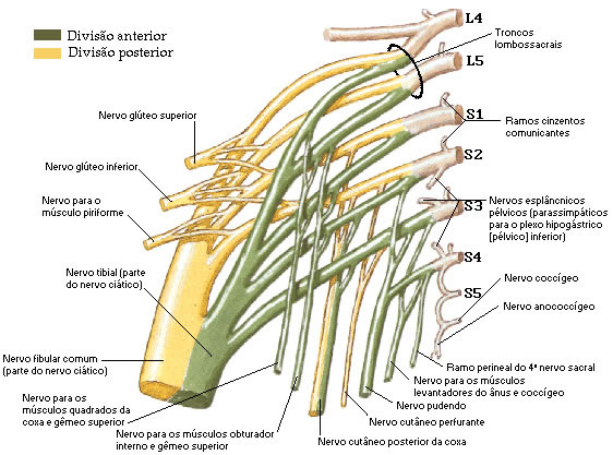
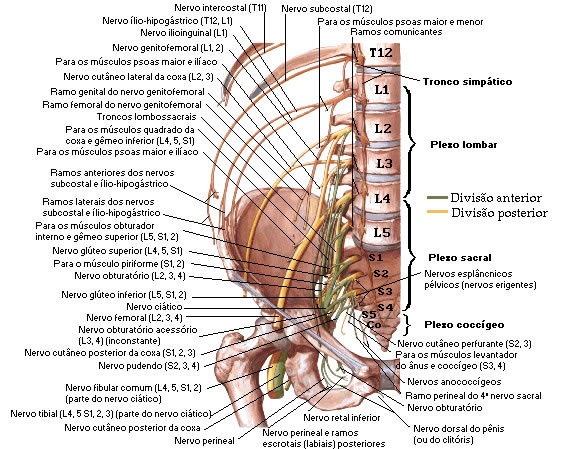
The ventral branches of the sacral and coccygeal spinal nerves form the sacral and coccygeal plexuses. The ventral branches of the four superior sacral nerves enter the pelvis through the anterior sacral foramina, the fifth sacral nerve penetrates between the sacrum and the coccyx, and the coccygeus below the coccyx.
Each ventral branch of the sacral nerves receives a gray communicating branch from a corresponding sympathetic ganglion. The efferent visceral branches leave the branches of the second to fourth sacral nerves as pelvic splanchnic nerves that contain the parasympathetic fibers, which reach minute ganglia in the walls of the pelvic viscera.
The organization of the sacral plexus is quite elementary and simple. The sacral plexus is formed by the lumbosacral trunk, ventral branches of the first to third sacral nerves, and part of the fourth, with the remainder of the latter joining the coccygeal plexus.
The anastomotic branch of L4 joins L5 to form the lumbosacral trunk. Then the lumbosacral trunk joins with S1 and then successively with S2, S3 and S4.
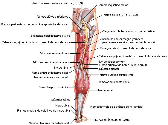
This nerve compact leaves the pelvis through the greater sciatic foramen. Shortly after crossing this foramen, the sacral plexus gives off its collateral branches and resolves into the terminal branch, which is the sciatic nerve. For the muscles of the gluteal region go the Superior Gluteal Nerves (L4, L5 and S1) and Inferior Gluteal Nerves (L5, S1 and S2). An important sensory branch is the Posterior Cutaneous Nerve of the Thigh , formed by S1, S2 and S3.
For the perineum, we have the Pudendo Nerve formed from S2, S3 and S4.
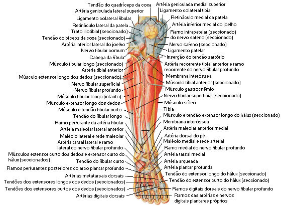
The Sciatic Nerve is the largest and most extensive nerve in the human body, as its fibers can go down to the toes. This nerve consists of two portions, which are the Common Peroneal Nerves (L4, L5, S1 and S2) and Tibial Nerves, formed by L4, L5, S1, S2 and S3. The common fibular nerve already in the popliteal fossa runs obliquely downward and laterally bifurcating into the superficial and deep fibular nerves.
The sacral plexus also leaves the nerves for the internal obturator muscle and superior twin muscle (L5, S1 and S2); for the piriformis muscle (S1 and S2); for the quadriceps muscle of the thigh and the lower twin muscle (L4, L5 and S1); for the levator ani, coccygeus and external anus sphincter muscles (S4); and the pelvic splanchnic nerve (S2, S3 and S4).
| LUMBOSACRAL PLEXUS |
 |
| Source: NETTER, Frank H.. Atlas of Human Anatomy. 2nd edition Porto Alegre: Artmed, 2000. |
| LUMBOSACRAL PLEXUS |
 |
Source: NETTER, Frank H.. Atlas of Human Anatomy. 2nd edition Porto Alegre: Artmed, 2000. |
| SUPERIOR GLUTEUS AND INFERIOR GLUTEUS NERVES |
 |
| Source: NETTER, Frank H.. Atlas of Human Anatomy. 2nd edition Porto Alegre: Artmed, 2000. |
| ISCHIATIC NERVE |
 |
| Source: NETTER, Frank H.. Atlas of Human Anatomy. 2nd edition Porto Alegre: Artmed, 2000. |
| FIBULAR NERVE |
 |
| Source: NETTER, Frank H.. Atlas of Human Anatomy. 2nd edition Porto Alegre: Artmed, 2000. |
| TIBIAL NERVE |
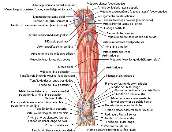 |
Source: NETTER, Frank H.. Atlas of Human Anatomy. 2nd edition Porto Alegre: Artmed, 2000. |
