Urinary system
The urinary system is constituted by Organs uropoetic organs, that is, responsible for preparing the urine and temporarily storing it until the opportunity to be eliminated to the outside. In the urine we find uric acid, urea, sodium, potassium, bicarbonate, etc.
This apparatus can be divided into secretory organs – which produce urine – and excretory organs – which are in charge of processing the drainage of urine out of the body.
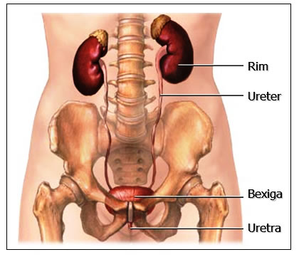
The urinary organs comprise the kidneys (2), which produce urine, the ureters (2) or ducts, which carry urine to the bladder (1), where it is retained for some time, and the urethra (1), through the which is expelled from the body.
In addition to the kidneys, the remaining structures of the urinary system function as a pipeline constituting the urinary tract pathways. These structures – ureters, bladder and urethra – do not modify urine along the way, rather they store and carry urine from the kidney to the external environment.
KIDNEY
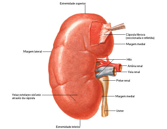
The kidneys are paired, bean-shaped organs located just above the waist, between the peritoneum and the posterior wall of the abdomen. Its color is red-brown.
The kidneys are situated on either side of the vertebral column, in front of the superior region of the posterior abdominal wall, extending between the 11th rib and the transverse process of the 3rd lumbar vertebra. They are described as retroperitoneal organs, as they are positioned behind the peritoneum of the abdominal cavity.
 The kidneys are covered by the peritoneum and surrounded by a mass of fat and loose areolar tissue. Each kidney is about 11.25 cm long, 5 to 7.5 cm wide, and just over 2.5 cm thick. The left is a little longer and narrower than the right. The weight of the adult male kidney varies between 125 to 170 g; in adult women, between 115 to 155 g. The right kidney is normally situated slightly below the left kidney because of the large size of the right lobe of the liver.
At the concave medial border of each kidney is a vertical cleft – the RENAL HILO – where the renal artery enters and the renal vein and pelvis leave the renal sinus. At the hilum, the renal vein is anterior to the renal artery, which is anterior to the renal pelvis. The renal hilum is the entrance to a space within the kidney.
The renal sinus, which is occupied by the renal pelvis, calyces, nerves, blood and lymph vessels, and a variable amount of fat.
External configuration of the kidneys:
Each kidney has two faces, two edges and two ends.
FACES (2) – Front and Back. Both are smooth, but the anterior is more domed and the posterior is flatter.
EDGES (2) – Medial (concave) and Lateral (convex).
EXTREMITIES (2) – Upper (Supra-Renal Gland) and Lower (at L3 level).
Internal configuration of the kidneys:
In a frontal cut through the kidney, two distinct regions are revealed: a smooth-textured reddish area called the renal cortex and a deep reddish-brown area called the renal medulla. The medulla consists of 8-18 cuneiform structures, the Renal Pyramids . The base (wider end) of each pyramid faces the cortex, and its apex (narrower end), called the renal papilla, points toward the hilum of the kidney. The parts of the renal cortex that extend between the renal pyramids are called renal columns.
Together, the renal cortex and renal pyramids of the renal medulla make up the functional part, or parenchyma, of the kidney. In the parenchyma are the functional units of the kidneys – about 1 million microscopic structures called NEPRONS . Urine, formed by the nephrons, drains into the large papillary ducts, which run along the renal papillae of the pyramids.

The ducts drain into structures called the Lesser and Greater Renal Calyces . Each kidney has 8-18 minor calyces and 2-3 major calyces. The lesser renal calyx receives urine from the papillary ducts of a renal papilla and transports it to a greater renal calyx. From the greater renal calyx, urine drains into the large cavity called the Renal Pelvis and then out through the Ureter to the urinary bladder. The renal hilum expands into a cavity in the kidney called the renal sinus.

nephrons
The nephron is the morphofunctional or urine-producing unit of the kidney. Each kidney contains about 1 million nephrons.
The shape of the nephron is peculiar, unmistakable, and admirably suited to its function of producing urine.
The nephron is made up of two main components:
1. Renal Corpuscle :
- Glomerular Capsule (Bowman's);
- Glomerulus – network of blood capillaries coiled inside the glomerular capsule
2. Renal Tubule :
- Proximal convoluted tubule;
- Loop of Nephron (of Henle);
- Distal convoluted tubule;
- Collecting tube.

Kidney Functions
The kidneys do the main work of the urinary system, with the other parts of the system primarily acting as passageways and storage areas. By filtering blood and forming urine, the kidneys contribute to the homeostasis of body fluids in a variety of ways.
Kidney functions include:
Regulation of the ionic composition of the blood;
- Maintenance of blood osmolarity;
- Blood volume regulation;
- Blood pressure regulation;
- Regulation of blood pH;
- Hormone release;
- Regulation of blood glucose level;
- Excretion of waste and foreign substances.
adrenal glands
The adrenal (adrenal) glands are located between the superomedial surfaces of the kidneys and the diaphragm. Each adrenal gland, surrounded by a fibrous capsule and a fat pad, has two parts: the adrenal cortex and medulla, both of which produce different hormones.
The cortex secretes hormones essential for life, while the medullary hormones are not essential for life. The adrenal medulla can be removed without causing life-threatening effects.
The adrenal medulla secretes two hormones: epinephrine (adrenaline) and norepinephrine. The adrenal cortex secretes steroids .
URETER:
There are two tubes that carry urine from the kidneys to the bladder.
 Small organs, the ureters are less than 6 mm in diameter and 25 to 30 cm in length.
Renal pelvis is the upper end of the ureter, located inside the kidney.
Descending obliquely downward and medially, the ureter travels in front of the posterior wall of the abdomen, then enters the pelvic cavity, opening at the ureter ostium located on the floor of the urinary bladder.
Due to its path, two parts of the ureter are distinguished: abdominal and pelvic. The ureters are capable of performing rhythmic contractions called peristalsis. Urine moves along the ureters in response to gravity and peristalsis.
BLADDER
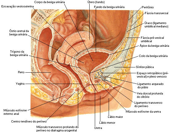
The urinary bladder functions as a temporary reservoir for storing urine. When empty, the bladder is located inferior to the peritoneum and posterior to the pubic symphysis: when full, it rises into the abdominal cavity.
It is a hollow, elastic muscular organ that, in men , lies directly anterior to the rectum. In women, it is in front of the vagina and below the uterus.
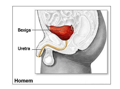
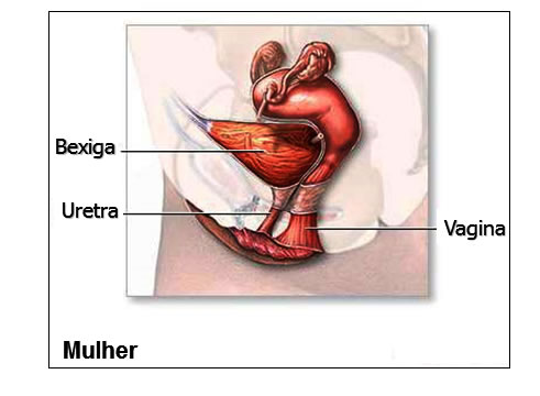
When the bladder is full, its inner surface is smooth. A triangular area on the posterior surface of the bladder does not show wrinkles. This area is called the bladder trigone and is always smooth. This triangle is limited by three vertices: the entry points of the two ureters and the exit point of the urethra. The trine is clinically important as infections tend to persist in this area.

The urinary bladder outlet contains the sphincter muscle called the internal sphincter, which contracts involuntarily, preventing emptying. Inferior to the sphincter muscle, surrounding the upper part of the urethra, is the external sphincter, which is controlled voluntarily, allowing resistance to the urge to urinate.
The average urinary bladder capacity is 700 – 800 ml; it is smaller in women because the uterus occupies the space immediately above the bladder.
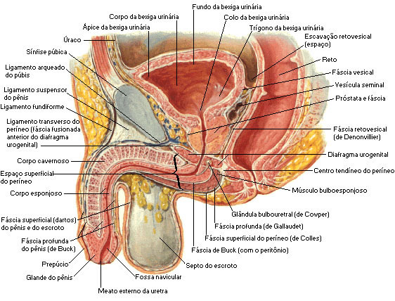
| MALE URINARY BLADDER |
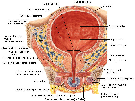 |
| Source: NETTER, Frank H.. Atlas of Human Anatomy. 2nd edition Porto Alegre: Artmed, 2000. |
| FEMALE URINARY BLADDER |
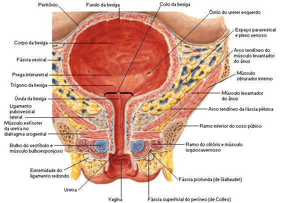 |
| Source: NETTER, Frank H.. Atlas of Human Anatomy. 2nd edition Porto Alegre: Artmed, 2000. |
The urethra is a tube that carries urine from the bladder to the external environment, being lined with mucosa that contains a large amount of mucus-secreting glands. The urethra opens to the outside through the external urethral orifice.
The urethra is different between the two sexes.
The male and female urethra differ in their path. In women, the urethra is short (3.8 cm) and is exclusively part of the urinary system. Its external ostium is located anterior to the vagina and between the labia minora. In men, the urethra is part of the urinary and reproductive systems. Measuring about 20 cm, it is much longer than the female urethra. When the male urethra leaves the bladder, it passes through the prostate and runs the length of the penis. Thus, the male urethra serves two purposes: it carries urine and sperm.
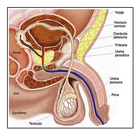 male urethra
The male urethra extends from the internal urethral orifice in the urinary bladder to the external urethral orifice at the end of the penis. It presents double curvature in the common state of relaxation of the penis. It is divided into three parts: the Prostatic , the Membrane and the Spongy , whose structures and relationships are essentially different. In the male urethra there is a tiny slit-shaped opening, an ejaculatory duct.

Female urethra
It is a narrow membranous canal extending from the bladder to the external orifice in the vestibule. It is placed dorsal to the pubic symphysis, included in the anterior wall of the vagina, and obliquely downwards and forwards; it is slightly curved, with the concavity directed forward. Its diameter, when not dilated, is about 6 mm. Its external orifice is immediately in front of the vaginal opening and about 2.5 cm dorsal to the glans of the clitoris. Many small urethral glands open into the urethra. The largest of these are the paraurethral glands, whose ducts empty directly into the urethral orifice.
