Skeletal System
Skeletal System Concept :
The skeletal system is made up of bones and cartilage.
I separated all the material in a Box for you. See more details now by clicking here.
Bones Concept:
Bones are whitish, very hard organs that, when joined to the others, through the joints or joints, constitute the Skeleton . It is a specialized form of connective tissue whose main feature is the mineralization (calcium) of its bone matrix (collagen fibers and proteoglycans).
Bone is a living, complex and dynamic tissue. A solid, highly specialized form of connective tissue that forms most of the skeleton and is the body's main supporting tissue. Bone tissue participates in a continuous dynamic remodeling process, producing new bone and degrading old bone.
Bone is made up of several different tissues: bone tissue, cartilaginous tissue, dense connective tissue, epithelial tissue, adipose tissue, nervous tissue, and various blood-forming tissues.
As for bone irrigation, we have Volkman's canals and Havers' canals. Bone tissue has no lymphatic vessels, only periosteal tissue has lymphatic drainage.
Havers' channels are a series of tubes around narrow channels formed by concentric lamellae of collagen fibers. This region is called compact bone or diaphysis. Blood vessels and nerve cells throughout the bone communicate by osteocytes (which emit cytoplasmic expansions that bring osteocytes into contact with each other) in gaps (spaces within the dense bone matrix that contain bone cells). This unique arrangement is conducive to mineral salt deposition, which gives bone tissue strength. It should also be noted that these channels run through the bone in the longitudinal direction, taking within its lumen, blood vessels and nerves that are responsible for the nutrition of the bone tissue. It causes blood vessels to pass through bone tissue.
Volkmann's canals are microscopic canals found in compact bone , are perpendicular to Haversian canals, and are a component of the Haversian system. Volkmann's canals can also carry small arteries throughout the bone. Volkmann's canals do not have concentric lamellae.
Inside the bone matrix are spaces called lacunae that contain bone cells called osteocytes. Each osteocyte has processes called canaliculi, which extend from the lacunae and join the canaliculi of neighboring lacunae, thus forming a network of canaliculi and lacunae throughout the mass of mineralized tissue.
Cartilage Concept :
It is an elastic form of semi-rigid connective tissue – it forms parts of the skeleton in which movement occurs. Cartilage has no blood supply of its own; consequently, your cells obtain oxygen and nutrients by long-range diffusion.
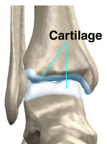
Articular Cartilage
Skeletal System Functions :
– Body support (support for the body)
– Protection of vital structures (heart, lungs, brain)
– Mechanical basis for movement
– Storage of salts (calcium, for example)
– Hematopoietic (continuous supply of new blood cells)
Number of Bones in the Human Body :
It is classic to admit the number of 206 bones.
Head=29 Skull=08 Face=14 Middle Ear Ossicles=3 neck=8 7 vertebrae hyoid Chest=37 24 ribs 12 vertebrae 1 sternum
abdomen=7 5 lumbar vertebrae 1 sacrum 1 coccyx | Senior Member=32 Shoulder Waist=2 arm=1 forearm=2 Hand=27
Lower Member=31 Pelvic Waist=1 Thigh=1 knee=1 leg=2 foot=26 |
Skeleton Division :
Axial Skeleton – Composed of the bones of the head, neck and trunk.
Appendicular Skeleton – Composed of the upper and lower limbs.
The union of the axial skeleton with the appendicular is made through the shoulder and pelvic girdles.

Bone Classification :
Bones are classified according to their shape into:
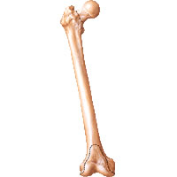
Long Bones: They are longer than they are wide and are made up of a body and two ends. They are slightly curved, which gives them greater strength. The slightly curved bone absorbs the mechanical stress of the body's weight at several points, in such a way that there is better distribution of the same. Long bones have their diaphysis formed by compact bone tissue and have a large amount of spongy bone tissue in their epiphyses. Example : Femur.
Short Bones: They are cube-like, having their lengths practically equal to their widths. They are composed of spongy bone, except on the surface, where there is a thin layer of compact bone tissue. Example : Carpal Bones.

Laminar (Flat) Bones: These are thin bones composed of two parallel layers of compact bone tissue, with a layer of spongy bone between them. Flat bones provide considerable protection and generate large areas for muscle attachment. Examples : Frontal and Parietal.
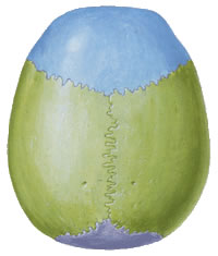
In addition to these three well-defined basic groups, there are other intermediates, which can be divided into 5 groups:
Elongated Bones: These are long bones, but flat and do not have a central canal. Example : Ribs.

Pneumatic Bones : They are hollow bones, with cavities filled with air and lined with mucosa (sinuses), with a small weight in relation to their volume. Example : Sphenoid.

Irregular Bones: They have complex shapes and cannot be grouped into any of the previous categories. They have varying amounts of cancellous and compact bone. Example : Vertebrae.

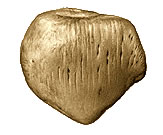
Sesamoid Bones : These are present within some tendons where there is considerable friction, tension and physical stress, such as the palms and soles. They can vary in size and number from person to person, they are not always completely ossified, typically only a few millimeters in diameter. Notable exceptions are the two patellas, which are large sesamoid bones present in almost all humans.
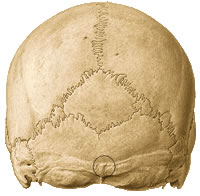
Structure of Long Bones :
The arrangement of compact and spongy bone tissue in a long bone is responsible for its strength. Long bones contain growth and remodeling sites, and structures associated with joints. The parts of a long bone are as follows:
Diaphysis : It is the long shaft of the bone. It consists mainly of compact bone tissue, providing considerable strength to the long bone.
Epiphysis : the flared ends of a long bone. The epiphysis of a bone articulates it, or joins it, to a second bone, in a joint. Each epiphysis consists of a thin layer of compact bone that lines the cancellous bone and is covered by cartilage.
Metaphysis : dilated part of the diaphysis closest to the epiphysis.


External Bone Configuration :
| BONE PROJECTIONS | |
| joint | non-articulating |
| - Head – condyle – facet | - Law Suit – Tubers – trochanter - Spine – Eminence – blades – Crests |
 |  |
| femur head | Transverse and Spinous Processes (Vertebrae) |
BONE DEPRESSIONS | |
| joint | non-articulating |
| – Cavities – acetabulum – fovea | – pits – Grooves – foramen - meatus - Breasts – Cracks - Channels |
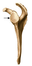 | 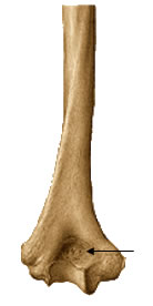 |
| Glenoid cavity (scapula) | Olecranon Fossa (Humer) |
Internal Configuration of Bones :
The differences between the two types of bone, compact and spongy or reticular, depend on the relative amount of solid substances and the amount and size of the spaces they contain. All bones have a thin superficial layer of compact bone surrounding a central mass of cancellous bone, except where the latter is replaced by a medullary cavity.
The compact bone of the body, or diaphysis, that surrounds the medullary cavity is the cortical substance. The architecture of cancellous and compact bone varies by function. Compact bone provides strength to support weight.
In long bones designed for stiffness and attachment of muscles and ligaments, the amount of compact bone is maximum, near the middle of the body where it is subject to bending. Bones have some elasticity (flexibility) and great rigidity.
Periosteum and Endosteum :
The periosteum is a dense, very fibrous connective tissue membrane that lines the outer surface of the diaphysis, firmly attaching to the entire outer surface of the bone, except for the articular cartilage. It protects the bone and serves as an attachment point for the muscles and contains the blood vessels that nourish the underlying bone.
The endosteum is located inside the medullary cavity of the bone, covered by connective tissue.
.jpg)
| Compact Bone Tissue | Spongy Bone Tissue |
| Contains few spaces in its rigid components. Provides protection and support and resists forces produced by weight and movement. Usually found in the diaphysis. | It makes up most of the bone tissue of short, flat, and irregular bones. Most are found in the epiphyses. |
